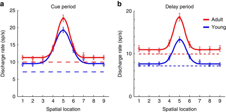
Figure 4. Tuning curve.
(a) Average activity (and s.e.m.) during the cue period of the ODR task in neurons recorded during the young (n=309) and adult stage (n=324). Locations have been rotated, so that the best location of each neuron is represented in location 5. Location 9 is the same as location 1. Solid lines represent best Gaussian fit of the population average. (b) Average activity (and s.e.m.) during the delay period of the ODR task in the same sample of neurons.

