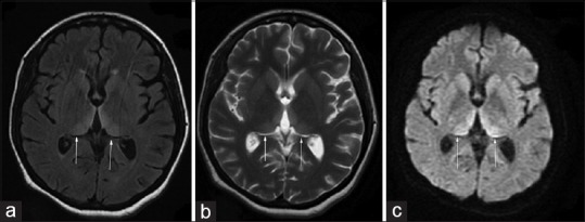
Figure 1.

(a) Magnetic resonance imaging of the brain T2 fluid-attenuated inversion recovery sequence showing bilateral medial thalamic hyperintensities (b) T2 sequence showing same findings (c) diffusion sequence showing high signal in bilateral medial thalamus
