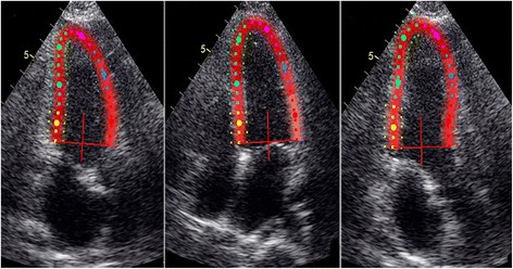
Fig 1.

Multiple-dotted lines in three myocardial layers at the three parasternal long-axis scans. The endocardial borders are delineated in the end-systolic frame of the images at the apical four-chamber, two-chamber, and long-axis planes. Subsequently, the myocardial wall is automatically defined with multiple chains of nodes for allowing assessment of longitudinal endocardial, mid-myocardial and epicardial strains
