
Fig. 1.
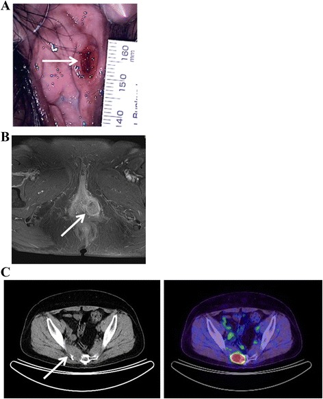
Physical examination and clinical imaging. a Ulcerative lesion on the inner side of the left labium minorum (arrow). b T1-weighted, contrast-enhanced magnetic resonance image revealing a 31 × 24 mm mass of moderate-to-high intensity in the left vulvar region (arrow); the mass does not involve the urethra or anus. c Positron emission tomography computed tomography revealing tumor metastases to the sacrum (arrow)