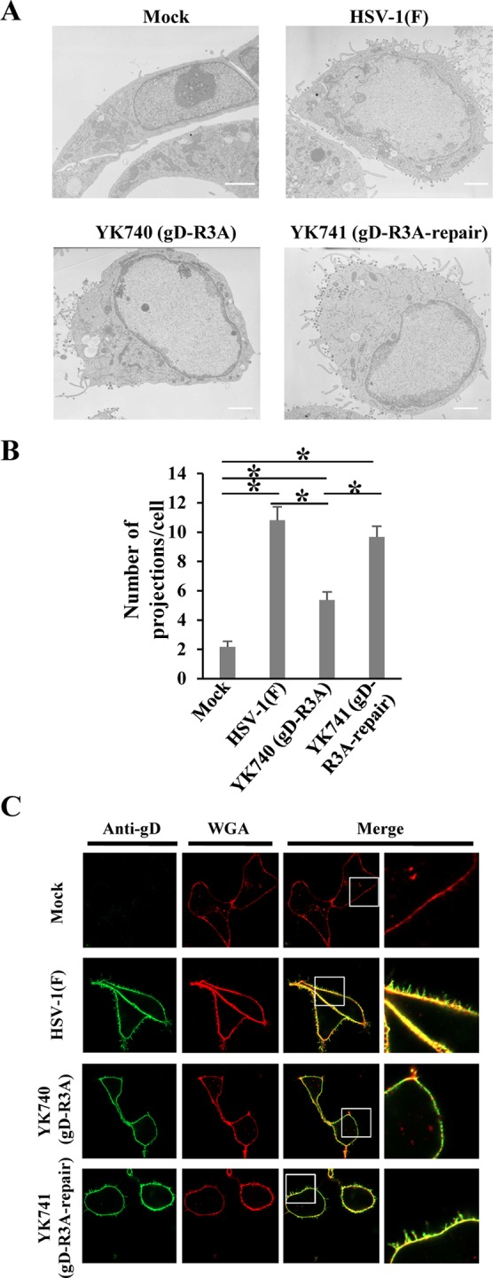
FIG 8.

Effect of mutations in the gD arginine cluster on projection formation at the plasma membrane in HSV-1-infected cells. (A) Vero cells were mock infected or infected with wild-type HSV-1(F), YK740 (gD-R3A), or YK741 (gD-R3A-repair) at an MOI of 5, fixed at 18 h postinfection, and examined by transmission electron microscopy. Bars, 2 μm. (B) Infected cells were examined by transmission electron microscopy as described in the legend to panel A, and the number of projections at the plasma membranes of 30 cells were quantitated. Statistical analysis was performed by one-way analysis of variance with the Tukey test. The asterisks indicate statistically significant differences, as follows: *, P < 0.05. (C) Vero cells were mock infected or infected with wild-type HSV-1(F), YK740 (gD-R3A), or YK741 (gD-R3A-repair) at an MOI of 3. At 18 h postinfection, infected cells were stained with WGA and anti-gD antibody without permeabilization and examined by confocal microscopy. Each image in the far right column is the magnified image of the boxed area in the image to its left.
