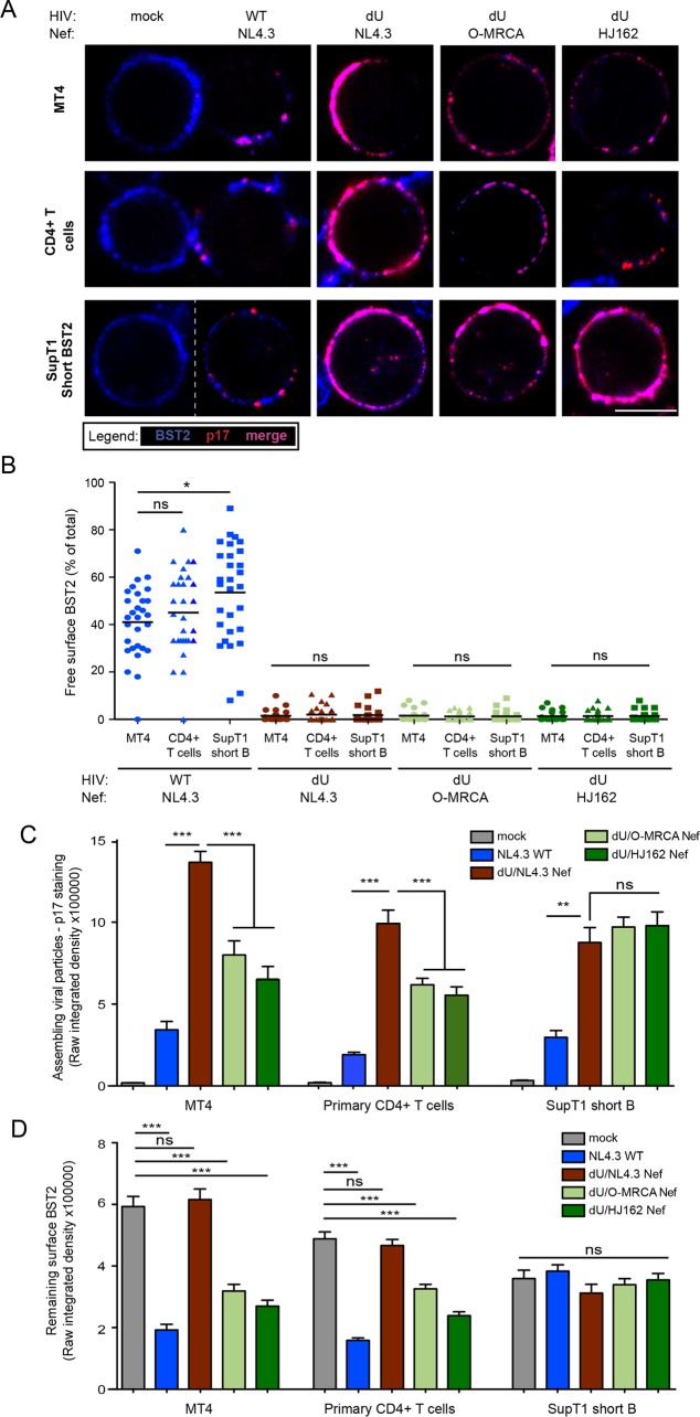
FIG 3.
O-Nef, unlike M-Vpu, cannot displace BST2 from sites of virus assembly. MT4 cells, primary CD4+ T cells, and SupT1 ShortBST2 cells were mock infected (mock) or infected with GFP-expressing NL4.3 WT and dU viruses expressing either NL4.3 Nef or O-Nefs, as indicated. (A) Cells were stained with anti-BST2 Abs, fixed, permeabilized, and then sequentially stained with anti-p17 Abs. An uninfected cell(mock) is shown next to WT-infected cells, as indicated. Bar = 10 μm. (B) The number of residual BST2 clusters not colocalizing with p17 (designated free BST2) per cell was calculated and expressed as the percentage of the total number of surface BST2 clusters. (C and D) The signal from assembling viral particles stained with p17 (C) or surface BST2 (D) was quantified by measuring the raw integrated signal density on manually selected cells using ImageJ software. Error bars indicate standard errors of the means after analysis of at least 50 distinct cells. One-way ANOVA with Bonferroni's multiple-comparison test was used (***, P < 0.001; **, P < 0.01; *, P < 0.05; ns, not significant [P > 0.05]).

