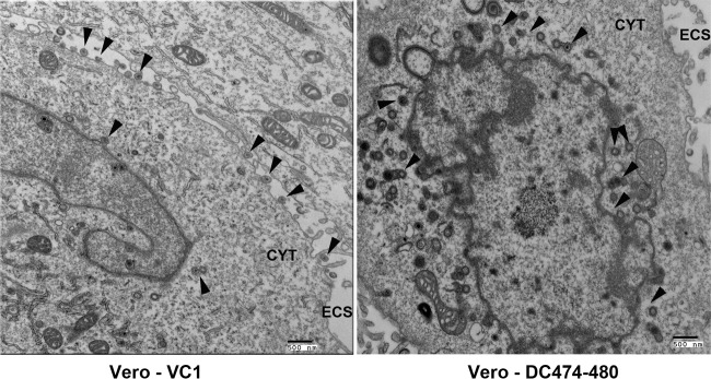
FIG 5.
Electron micrographs of Vero cells infected with VC1 or DC474-480 viruses. Vero cells were infected with either VC1 or DC474-480 viruses at an MOI of 5 and visualized by electron microscopy after 24 hpi. Arrowheads indicate the presence of enveloped virions in the periphery of VC1-infected cells and capsids within the cytoplasm of DC474-480-infected cells. The cytoplasm (CYT) and extracellular space (ECS) are indicated.

