FIG 7.
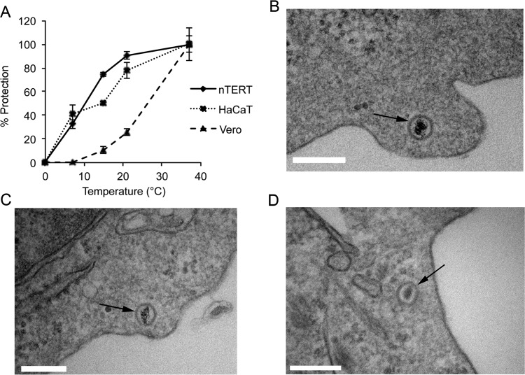
HSV-1 enters keratinocyte cells at low temperatures. (A) Confluent monolayers of nTERT, HaCaT, or Vero cells were infected with 200 PFU of HSV-1 per well of a 6-well plate on ice for 1 h. The inoculum was removed and replaced with medium at the temperature indicated and incubated at that temperature for 1 h before treatment with low-pH buffer for 1 min. After treatment, cells were washed and overlaid with medium containing 1% human serum. The infection was allowed to continue for 2 days, when plaques were fixed, stained, and counted. The means ± standard errors of the data are given from one representative experiment (n > 3). Data are displayed as the percentage of virus protected (% protection) at each temperature, where the plaque count at 37°C was taken as 100%. (B to D) Monolayers of nTERT cells seeded in 60-mm dishes were prechilled on ice and infected with 100 PFU per cell of gradient-purified HSV-1 on ice for 1 h. Cells were then shifted to 7°C for 60 min before fixing on ice for TEM processing. Vertical sections were analyzed, and representative images are shown with arrows identifying intracellular capsids. Scale bar, 200 nm.

