
FIG 3.
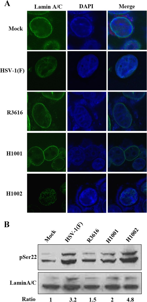
(A) Effect of γ134.5 variants on reorganization of lamin A/C. HeLa cells were mock infected or infected with HSV-1(F), R3616, H1001, or H1002 (5 PFU/cell). At 16 h postinfection, cells were processed and stained with DAPI (4′,6′-diamidino-2-phenylindole) and anti-lamin A/C antibody. Cells were visualized, and images were captured with confocal microscopy. (B) Effect of γ134.5 variants on phosphorylation of lamin A/C. HeLa cells were infected as for panel A. Lysates of cells were prepared and processed for Western blot analysis with antibodies against lamin A/C and phosphorylated lamin A/C (serine 22), respectively. Phosphorylated lamin A/C (serine 22) and lamin A/C were quantitated using Image J software. Phosphorylation of lamin A/C is presented as a ratio to lamin A/C, with normalization to mock infection.