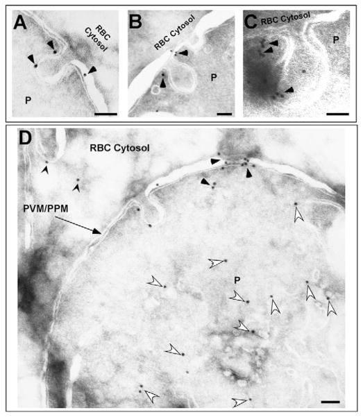
Fig. 10.
Immunoelectron microscopy depicting the localization of Pfactin in JAS-treated PE. (A–D) Representative electron micrographs depicting localization of Pfactin in JAS-treated trophozoite PE. (A–C) Pfactin associated with the neck and body of the cytostome (black arrowheads). (D) Pfactin is specifically labeled with the anti-actin antibody in the PVM/PPM (black arrowheads) and the parasite cytosol (white arrowheads). In addition, some Pfactin is present in the erythrocyte cytosol (pointed black arrowheads). Scale bar: 100 nm.

