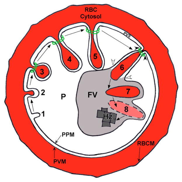
Fig. 13.
A new model for hemoglobin transport to the FV. (Steps 1–3) Cytostome formation. (1) The PVM invaginates after which (2) the PPM invaginates. (3) A double-membrane electron-dense collar forms around the cytostome. (Steps 4–6) Cytostome maturation. (4) The cytostome continues to fill with red blood cell cytosol and hemoglobin, and (5) elongates to appose the FV. (Steps 6–8) Hemoglobin deposition and degradation in the FV. (6) Fusion occurs between the matured cytostome and the FV (white arrowheads), while, simultaneously, the cytostome pinches off from the PVM and PPM (black arrowheads), resulting in the release (7) of a single-membrane-bound hemoglobin-filled vesicle into the FV lumen. During Step 6 (white arrowheads) content mixing occurs between the FV lumen and the PVS. (8) The membrane of this vesicle is degraded by FV-resident lipases, while the hemoglobin is degraded by FV-resident proteases. The resulting heme is polymerized into hemozoin (Hz). P, parasite; PPM, parasite plasma membrane; PVM, parasitophorous vacuolar membrane; PVS, parasitophorous vacuolar space; RBC, red blood cell; RBCM, red blood cell membrane.

