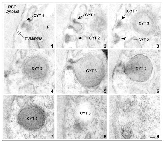
Fig. 2.
Hemoglobin-containing compartments are contiguous with the cytostome. Electron micrographs of serial thin sections (sections 1–9) from a representative trophozoite stage PE. Three cytostomes, CYT 1, CYT 2 and CYT 3, are apparent. CYT, cytostome; P, parasite; RBC, red blood cell; PPM, parasite plasma membrane; PVM, parasitophorous vacuolar membrane. Scale bar: 100 nm.

