
Fig. 1.
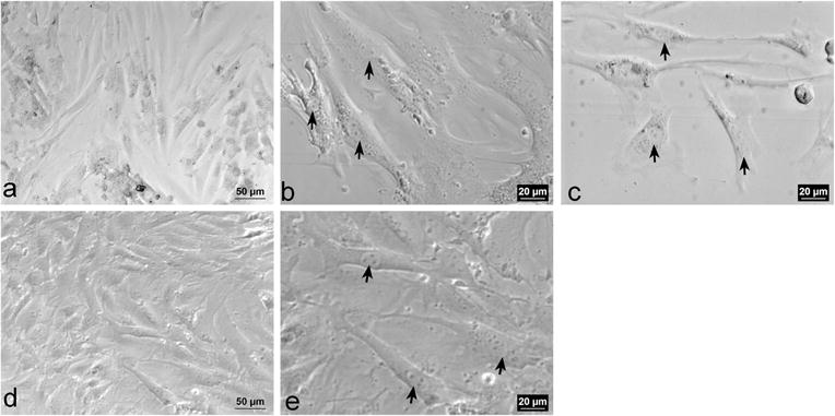
Representative phase-contrast images of P1 VSMCs. a–c Represent P1 VSMCs a Confluent monolayer of P1 VSMCs (×20). b and c Spindle-shaped VSMCs with phase dense cytoplasm and oval nuclei with two nucleoli (arrows, ×40). d–e Represent P2 VSMCs. d Confluent monolayer of P2 VSMCs (×20). e Spindle-shaped VSMCs with oval nuclei containing 3–5 nucleoli (arrows, ×40)