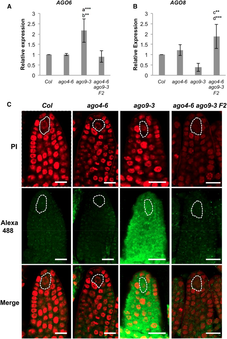
Figure 4.
The expression of AGO6 and AGO8 is affected by loss-of-function mutations in ago4 or ago9. (A) Expression of AGO6 in developing gynoecia containing premeiotic ovules. (B) Expression of AGO8 in developing gynoecia containing premeiotic ovules. The comparative 2−ΔΔCt method was used for determining the relative level of gene expression as compared to wild type, using ACTIN2 as internal control (Czechowski et al. 2004). Each histogram represents the mean of three biological replicates and shows the corresponding SD. Letters indicate pairwise results of two-tailed Fisher’s exact tests used to estimate statistical significance of possible differences between genotypes: a, comparison to Col; b, comparison to ago4-6 ago9-3 F2; c, comparison to Col; d, comparison to ago9-3. ** P < 0.01, *** P < 0.001. (C) Whole-mount immunolocalizations showing the expression of AGO6 in wild-type and mutant premeiotic ovules. Alexa 488 fluorescence (green) denotes the localization of the antibody raised against AGO6; samples were counterstained with propidium iodide (red). Bar, 15 µm.

