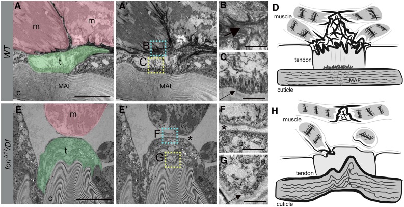
Figure 5.
Loss of Fon alters cuticle integrity, tendon cell cytoarchitecture, and ECM accumulation. (A–C and E–G) TEMs of an indirect muscle–tendon attachment site in WT (A–C) or fonΔ17/Df mutants (E–G). (A and A′) Muscles (m; pink) are interlaced with tendon cells (t; green) at control MTJs. The insets correspond to high magnification images in B (cyan) and C (yellow). (B) An electron-dense ECM matrix is observed between the sarcolemma and tendon cell membranes (large arrow). (C) Apical junctions present at the base of tendon cells (small arrow) are associated with MAFs (A and A′) that extend into the cuticle. (D) Generalized schematic representation of WT muscle attachment. (E and E′) The detached muscle (m; pink) in this fon mutant remains close to the tendon cell (t; green), which is attached to a highly convoluted cuticle (c). The insets correspond to close-up images in F (cyan) and G (yellow). (F) There is a loss of electron-dense ECM material and membrane interdigitation between the muscle and tendon cell (asterisk). (G) Note the absence of tendon cell junctional complexes at the base of the tendon cell (G, small arrow). (H) Illustration reveals the dramatic loss of muscle attachment, ECM accumulation, and morphological abnormalities associated with mutations in fon. Bars, 10 µm for A and E; 1 µm for B, C, F, and G.

