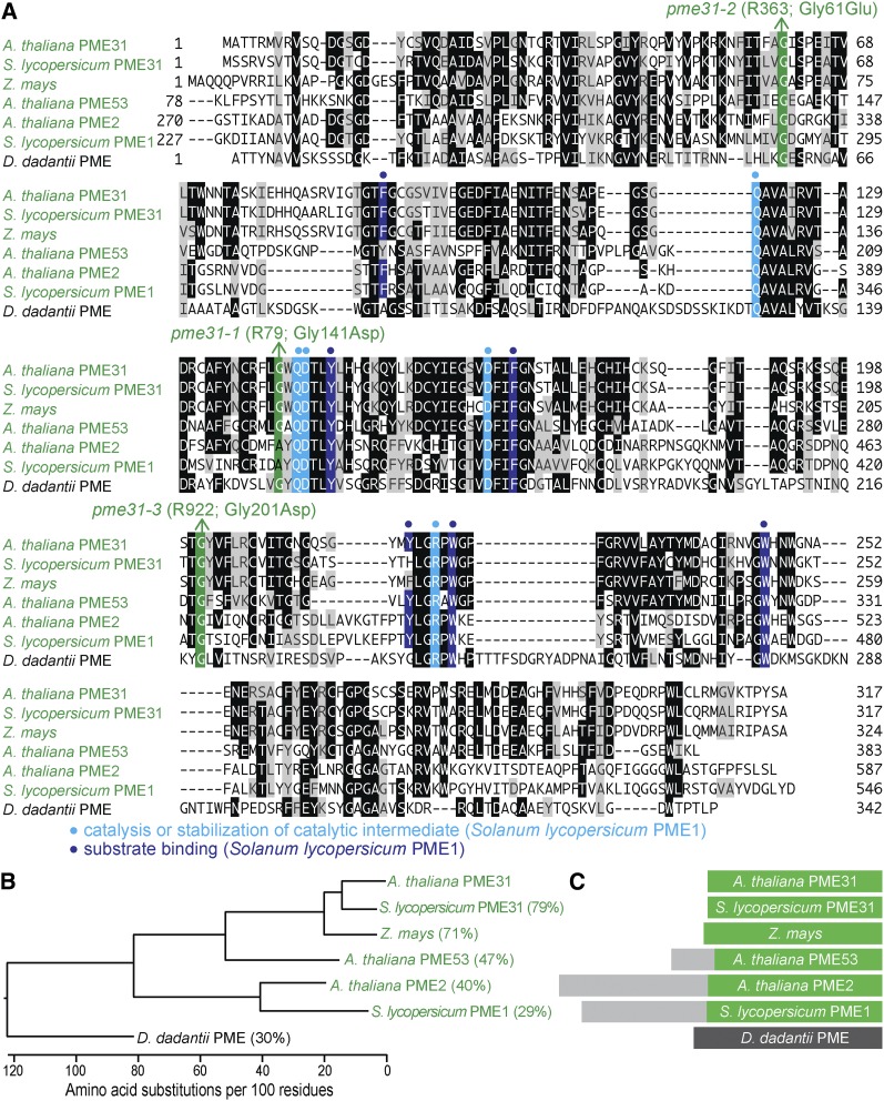
Figure 15.
PME31 is closely related to other PME proteins and lacks a predicted signal peptide. (A) PME31 was aligned with related proteins (Table S4); residues are shaded when identical (black or colors) or chemically similar (gray) in at least three sequences. Residues important for catalysis or stabilization of a catalytic intermediate (light blue background and dots) and for substrate binding (dark blue background and dots) in S. lycopersicum PME1 are indicated. Mutations identified in this work are denoted (green). (B) Phylogenetic tree of PME proteins constructed using ClustalW on Lasergene MegAlign (DNAStar). (C) Diagram of PME proteins, showing sections of the protein that align (green and dark gray) and the N-terminal extensions present only in some PME proteins (light gray) that target these PME enzymes for secretion to the cell wall (Dedeurwaerder et al. 2009).

