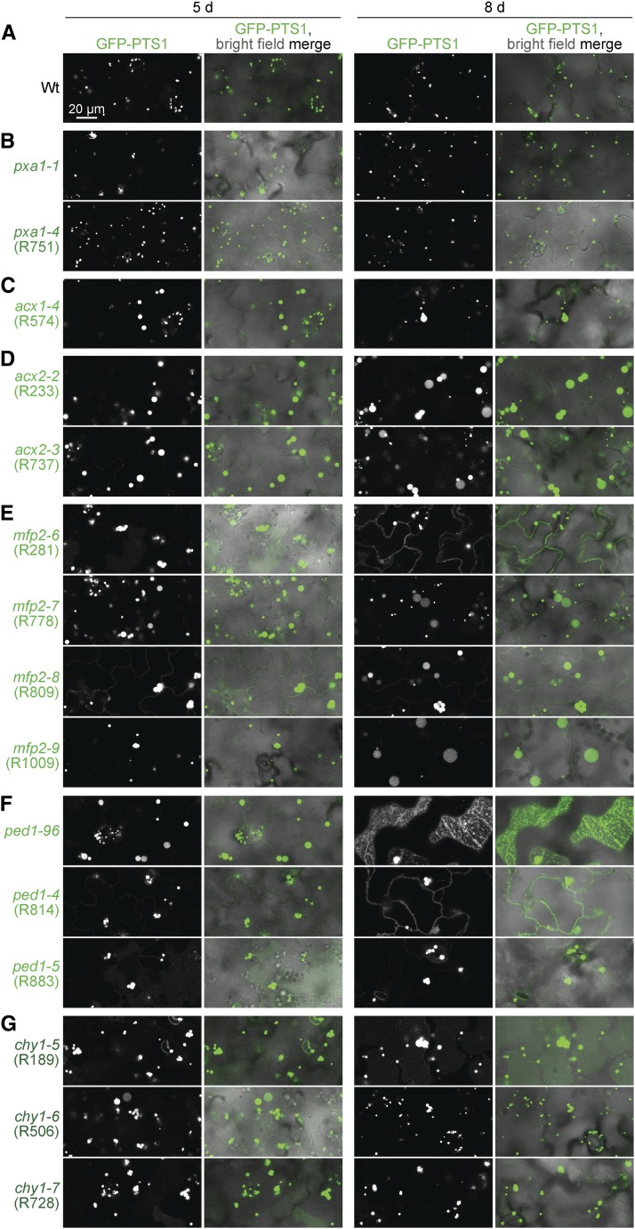
Figure 9.
A subset of fatty acid β-oxidation mutants display enlarged peroxisomes and matrix protein import defects. (A–G) Wild type (A) and pxa1 mutants (B) display small peroxisomes, whereas mutations in ACX1 (C), ACX2 (D), MFP2 (E), PED1 (F), and CHY1 (G) lead to enlarged peroxisomes and some GFP-PTS1 mislocalization to the cytosol. Confocal micrographs were taken of cotyledon epidermal cells of 5- or 8-day-old seedlings expressing peroxisomally targeted fluorescence (GFP-PTS1; white in left panels; green in merged images). Data are representative of three replicates. Bar, 20 µm.

