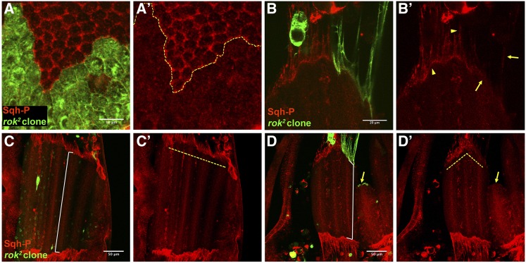
Figure 2.
Drok loss of function in tendon cells results in diminished Sqh phosphorylation and disturbs muscle morphogenesis. (A, A’) Drok2 mutant clone of notum epithelial cells (27 hAPF) marked with CD8-GFP displays diminished levels of monophosphorylated Sqh (Sqh-P) at the apical side. (B, B’) Drok2 mutant tendon cell processes and myotendinous junction display diminished levels of Sqh-P (arrows), cf. wild type tendon extension and myotendinous junction (arrowheads). (C–D’) Confocal projections of DLMs at 27 hAPF of an animal carrying a Drok2 clone at the right side (D, D’). DLMs attached to wild type tendon cells display a regular anterior edge (dashed yellow line) (C’), while DLMs attached to a group of Drok2 tendon cell processes display irregular anterior edge (dashed yellow line) (D’) and display detachment of the epithelia (arrow). Note that myotubes attached to Drok2 tendon processes are shorter than the contra lateral muscles, cf. white guide of (C) and (D).

