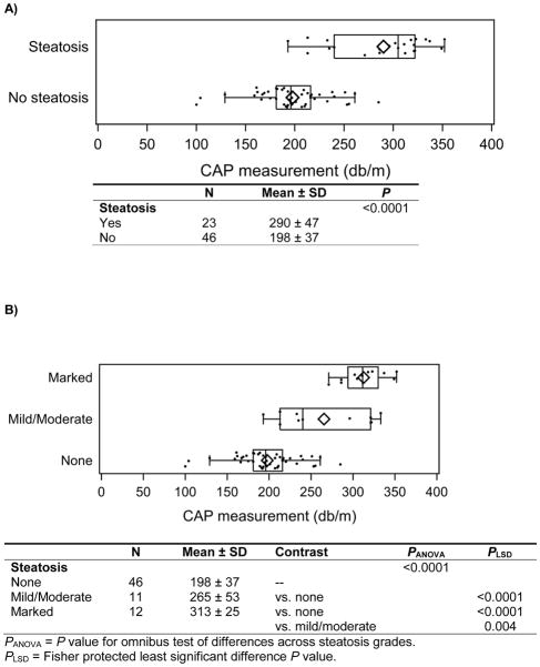
Figure 1.
A) CAP measurement by presence of biopsy-confirmed steatosis (n=69). The box-whisker plots describe the distribution of CAP measurements, with the left and right edges of each box corresponding to the lower and upper quartiles, the vertical line and diamond within representing the median and mean respectively, and the whiskers extending up to 1.5 times the interquartile range (i.e. box width). B) Distribution of CAP measurement by degree of biopsy-confirmed steatosis (n=69). The box-whisker plots describe the distribution of CAP measurements, with the left and right edges of each box corresponding to the lower and upper quartiles, the vertical line and diamond within representing the median and mean respectively, and the whiskers extending up to 1.5 times the interquartile range (i.e. box width).

