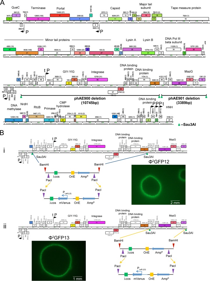
FIG 2.
Generation of DS6A fluorescent reporter phages. (A) The two nonessential regions in DS6A identified after deletion mutagenesis site mapping of all the recombinant DS6A phages are shown. The first deletion of ∼11 kb spanned genes gp42 to gp65, and the resulting phage was named phAE900. The second deletion mapped to an adjacent 3.4-kb region spanning gp66 to gp72, and the resulting phage was named phAE901. (B) The pYUB328 plasmid inserted in phAE900 and phAE901 was replaced by the pYUB1552 plasmid, resulting in Φ2GFP12 (i) and Φ2GFP13 (ii), respectively. Comparable expression of mVenus from the Pleft (L5) promoter resulted in plaque boundaries of similar fluorescence intensities after electroporation of Φ2GFP12 and Φ2GFP13 into M. tuberculosis. Genome maps were generated using Phamerator (53), with rightward- and leftward-transcribed genes shown as colored boxes above and below the genome, respectively. Genes are sorted into phamilies according to sequence similarity (37); the pham assignment number is shown above or below each gene with the number of phamily members in parentheses. Genes are colored according to their pham assignment, and white-colored genes are those that do not have homologues among other mycobacteriophages.

