Figure 1.
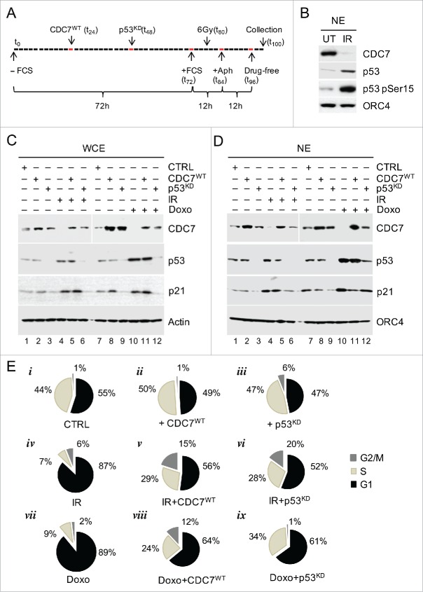
DNA Damage Induces G1 Arrest via p53-dependent Down-regulation of CDC7. (A) Schematic presentation of the synchronisation protocol (see Methods).55 p53 siRNA was introduced 12h prior and irradiation (IR) 8h after the end of serum starvation. Cells were collected for analysis 4h after release from the second Aphidicolin block. (B) Immunoblotting analysis of the nuclear extract (NE) from untreated (UT) and IR cells probed with the indicated antibodies. Orc4 was used as a loading control. (C) Immunoblotting analysis of whole cell extracts (WCE) from synchronised IMR90 cells treated with either IR (6 Gy) or with Doxorubicin (Doxo) independently or in conjunction with either CDC7 overexpression (CDC7WT) and/or p53 silencing (p53KD), probed with the indicated antibodies. β-actin was used as a loading control. (D) Immunoblotting analysis of nuclear extracts (NE) from synchronised IMR90 cells treated with either IR (6 Gy) or Doxo independently or in conjunction with either CDC7 overexpression (CDC7WT) and/or p53 silencing (p53KD), probed with the indicated antibodies. Orc4 was used as a loading control. (E) FACS analyses are shown as pie charts to demonstrate that the G1 arrest induced by IR or Doxo can be abrogated by either p53 knockdown (p53KD) or ectopic expression of CDC7 (CDC7WT), as demonstrated by the resumption of cell cycle progression into the S-phase.

