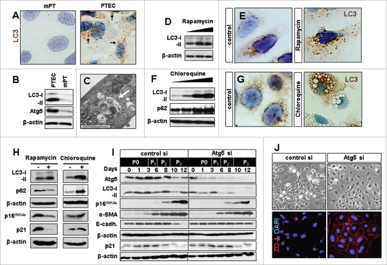
Figure 1.

Experimental modification of baseline autophagy influences senescence marker expression and epithelial phenotype in renal primary tubular epithelial cells (PTEC). In vitro culturing of PTEC induces autophagy. (A) LC3 staining in mouse proximal tubular cells (mPT) cells and PTEC showing LC3 punctae (arrows) which are localized on the autophagosomal membrane during autophagy induction. (B) Representative LC3 immunoblots in PTEC as compared to mPT cells. (C) Electron microscopy image of PTEC with autophagic vacuoles representing baseline autophagy. (D) LC3 immunoblot in PTEC treated with increasing concentrations of rapamycin. (E) Immunocytochemistry for LC3 punctae upon autophagy induction with rapamycin. (F) Representative immunoblots for LC3 and p62 and (G) immunocytochemistry for LC3 aggregates and vacuole formation in PTEC upon autophagy inhibition with increasing concentrations of chloroquine. (H) Immunoblots for LC3, p62, p16INK4a and p21 at day 12 in PTEC treated with rapamycin and chloroquine. (I) Immunoblots for Atg5, LC3, p16INK4a, α-smooth muscle actin (α-SMA), E-cadherin (E-cadh.) and p21 in Atg5 or control siRNA treated PTEC harvested at indicated time points and passages (P0-P3). (J) Representative pictures of day 12 PTEC transfected with control or Atg5 siRNA in bright field and immunohistochemistry for epithelial marker ZO-1. Original magnification x 630 in A, E, and G and x 400 in J. Scale bar 1 µm in C. n = 3.
