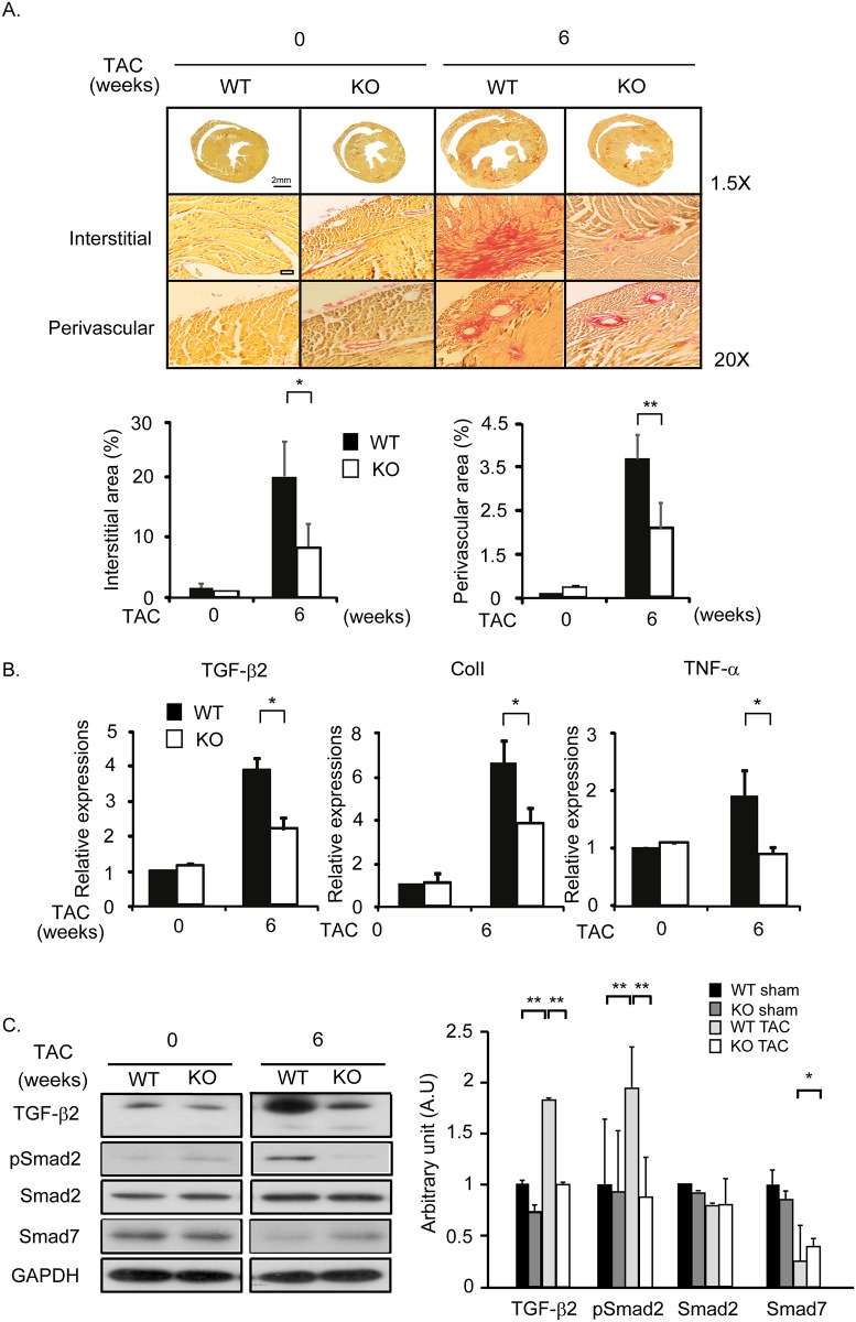
Fig 2. CF is attenuated in cytl1 KO mice.
WT and cytl1 KO mice were subjected to TAC for 6 wks and the extent of fibrosis in the heart was analyzed. (A) Picrosirius staining of heart cross-sections from WT and cytl1 KO mice subjected to TAC. Fibrotic areas in the interstitial and perivascular areas were quantified using MetaMorph software (right panels). (B) Quantification of the mRNA levels of several fibrotic markers (TGF-β2, collagen 1 and TNF-α) by qRT-PCR. (C) Activation of the TGF-β signaling pathway was investigated by western blotting. GAPDH served as the loading control. n = 3–5 for each experimental group. *p < 0.05, **p < 0.01.

