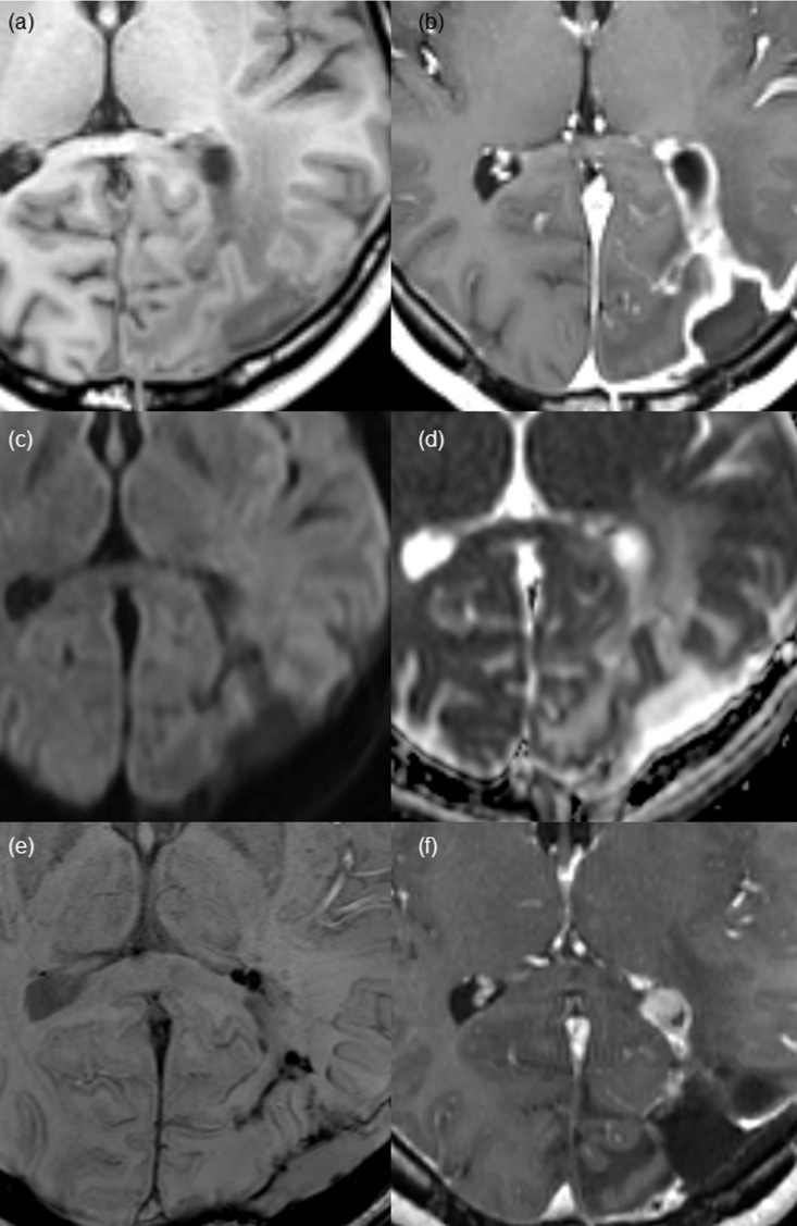
Fig 4. Nodular wall enhancement pattern found on post-CCRT MR imaging in a 36-year-old female patient after gross total resection of glioblastoma.
(a and b) Pre- and post-contrast enhanced T1 WI demonstrates a nodular enhancement of 7 mm in the diameter at the anterior margin of the surgical cavity with thick wall enhancement. No demonstrable diffusion restrictions on (c) DWI or (d) ADC map in the enhancing portion. (e) Irregular dark signal intensity is shown along the surgical cavity on SWI, suggestive of hemorrhage. (f) At 15-month follow-up MR imaging, a measurable nodular enhancing lesion appears in the same portion of the surgical cavity, which is pathologically proven recurred glioblastoma.

