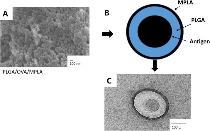
Fig 1. Characterization of PLGA NPs.
(A) Morphological analysis of PLGA NP/OVA/MPLA by scanning electron microscopy (SEM). (B) Schematic representation of the PLGA/OVA/MPLA NP and (C) SEM of a single PLGA NP/OVA/MPLA. Scale bar = 100 nm and 100 μ. PLGA: poly-lactic-co-glycolic acid, MPLA: Monophosphoryl Lipid A, OVA: ovalbumin

