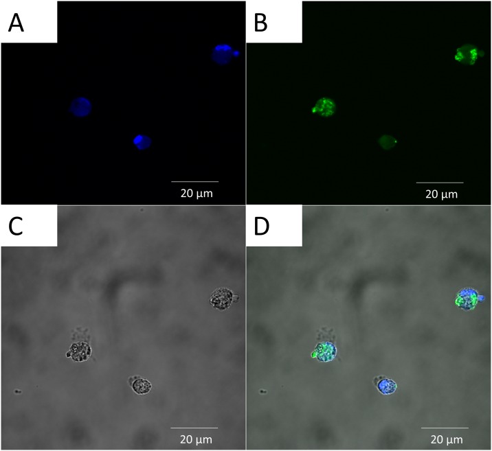
Fig 6. Cellular localization of PLGA/OVA-FITC/MPLA NP uptake by canine macrophages and evaluated by confocal laser scanning microscopy after 2 hours of incubation.
Day 7 canine macrophages cultures (1x10⁶) were incubated with 50 μg/mL of PLGA/OVA-FITC/ MPLA NPs. After 2 hours, macrophages were harvested and analyzed by confocal microscopy. One representative experiment is shown. (A) Nucleus of macrophages stain blue by DAPI. (B) PLGA/OVA-FITC/MPLA NP stain green. (C) Canine macrophages. (D) Merge of A&B demonstrating internalization of particles. Scale bar = 20 μm. DAPI = 4',6-Diamidino-2-Phenylindole, Dihydrochloride; FITC = Fluorescein isothiocyanate. Similar data were obtained in three independent experiments.

