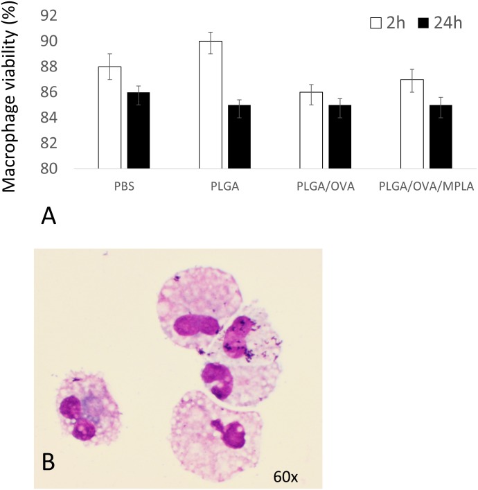
Fig 7. Evaluation of canine macrophage viability incubated with PLGA NP formulations.
Day 7 canine macrophages cultures (1x10⁶) were incubated with 50 μg/mL of different NP formulations for 2 hours or 24 hours at 37°C. Macrophages were then stained with Trypan Blue and evaluated microscopically. (A) Cells that take the stain have impaired membranes and are considered dead cells. (B) Day 7 canine macrophages (1x10⁶), were cytocentrifuged and evaluated under light microscopy for evidence of pyknosis or cellular necrosis. Similar data were obtained in three independent experiments.

