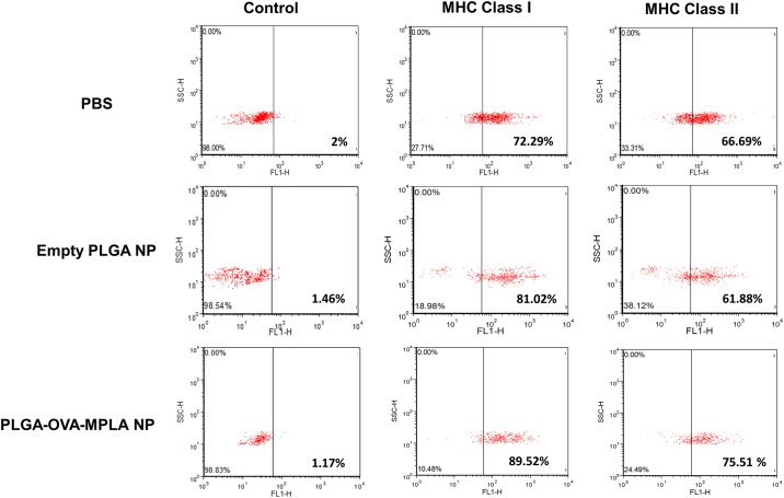
Fig 9. Flow cytometric evaluation of MHC-I and MHC-II phenotype in canine macrophages exposed to PLGA NP preparations.
Day 7 canine macrophages (1x10⁶) were incubated with 50 μg/mL of PLGA NP or PLGA/OVA/MPLA NP. After 24 hours, nonadherent cells were harvested and analyzed by flow cytometry. FSC (forward scatter) and SSC (side scatter) profiles of PBS and PLGA NP-incubated with canine macrophages are shown. One color flow cytometry dot plots gated indicating the phenotype of macrophages incubated with PLGA NPs. One representative experiment is shown.

