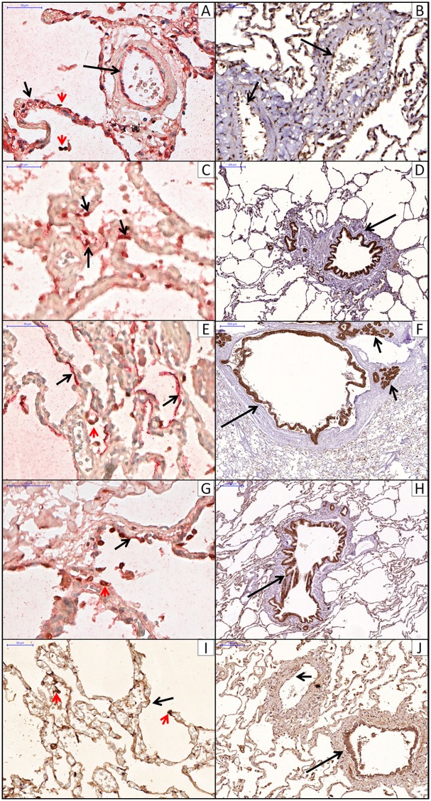
Fig 2. Immunohistochemistry localization of aquaporins, Na-K-ATPase channel and ENaC channel.
A: AQP1 in alveolar (short arrow) and arteriolar (long arrow) endothelial cells. Double staining for AQP1 (red) and PII cels (TTF-1, brown, red arrowhead). B: AQP1 in vascular endothelial cells (arrow). C: AQP3 in PII cells (arrows). Double staining for AQP3 (red) and PII cels (TTF1, brown). D: AQP3 in epithelial bronchiolar cells (arrow). E: AQP5 in PI cells lining the alveolar septum (arrow). Double staining for AQP5 (red) and PII cels (TTF1, brown, red arrowhead). F: AQP5 in epithelial bronchiolar cells (long arrow) and submucosal glands (short arrow). G: Na-K-ATPase channel in PI (arrow) and PII (red arrowhead) cells. Double staining for Na-K-ATPase channel (red) and PII cels (TTF1, brown). H: ENaC channel in PI cells lining the alveolar septum (arrow) and PII cells (red arrowhead). I: ENaC channel in epithelial bronchiolar cells (long arrow) and vascular endotelial cells (short arrow).

