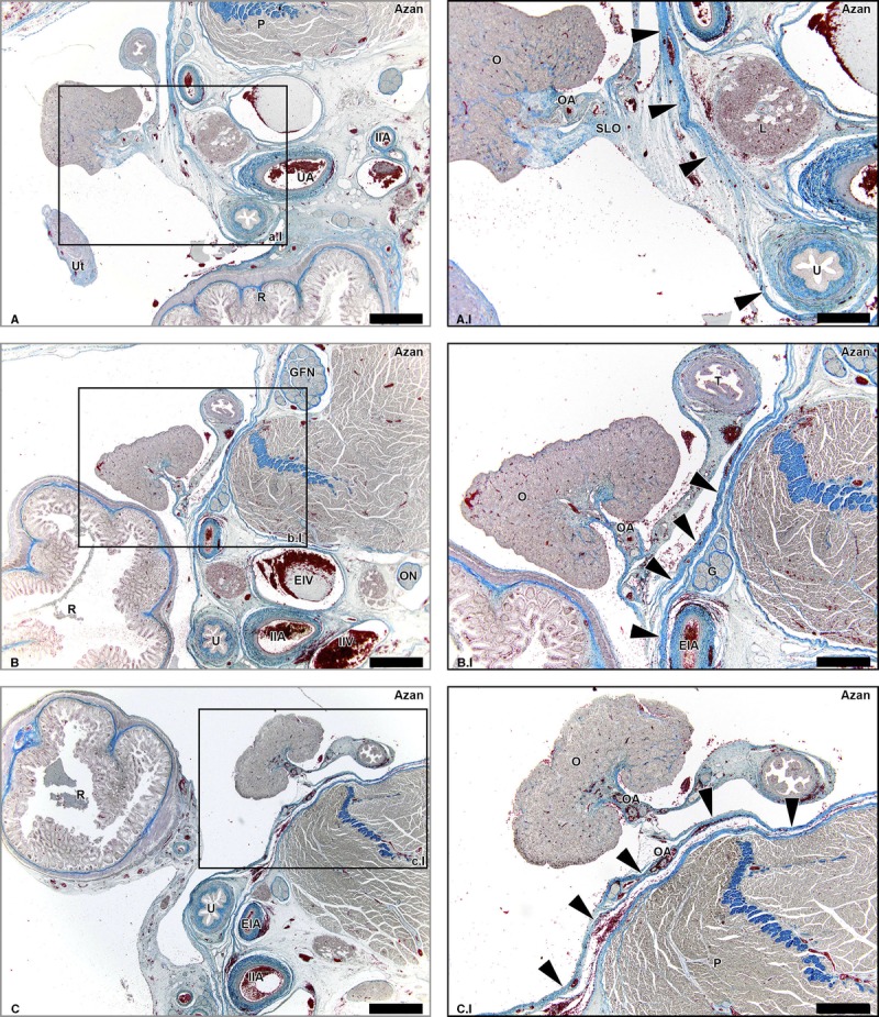
FIGURE 2.

Suspensory ligament of the ovary (infundibulopelvic ligament) and retroperitoneum. Transverse azan-stained sections of the fetus aged 15 weeks showing collagen (blue) and cell nuclei (purple) at consecutive superior levels (A-C) The SLO does not form a direct continuity with the retroperitoneum as the peritoneum (arrowheads) forms a solid border. Lymph vessels draining via the SLO do not cross this border, but run along the OA and shift to the retroperitoneal compartment when the OA does (window C.I). P indicates psoas major muscle; Ut, uterus; UA, umbilical artery; IIA, internal iliac artery; R, rectum; O, ovary; U, ureter; EIA, external iliac artery; L, lymph nodes along the EIA; GFN, genitofemoral nerve; EIV, external iliac vein; ON, obturator nerve; IAV, internal iliac vein; T, uterine tube; G, genital branch of GFN. Scale bar overview, 1 mm; scale bar detail, 500 μm.
