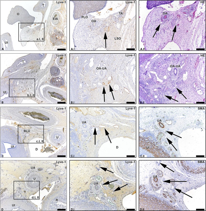
FIGURE 3.

Lymph vessels draining the ovary via the ovarian ligament and the broad ligament of the uterus. Transverse sections of the fetus aged 14 weeks showing the lymphatic drainage pathway of the ovary (O) via the proper ligament of the ovary (PLO) and broad ligament of the ovary (BLO) at consecutive inferior levels (A-D). The arrows in the Lyve-1–stained sections show the lymph vessels running along the OA, merging eventually with the supraureteral pathway that follows the course of the uterine artery (UA). Ut indicates uterus; T, uterine tube; EIA, external iliac artery; L, lymph nodes along the EIA; TA, tubal branches of OA; OA-UA, anastomosis of OA and UA; R, rectum; U, ureter; D, Douglas’ pouch. Scale bar overview, 500 μm; scale bar detail, 200 μm.
