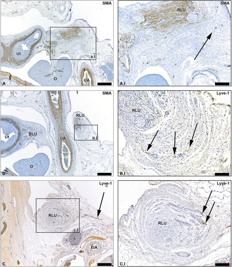
FIGURE 4.

Lymph vessels draining the ovary via the round ligament of the uterus. Transverse sections of the fetus aged 14 weeks showing lymph vessels (arrows) that drain the ovary (O) via the round ligament of the uterus (RLU, ligament teres uteri) at consecutive inferior levels (A-C). Note the presence of smooth muscle fibers in the RLU (brown stain in the SMA-stained sections). At an inferior level, the RLU passes under the umbilical artery (UA) as can be seen in window B. The RLU runs to the inguinal region and its lymph vessels eventually fuse with inguinal lymph vessels. Ut indicates uterus; O, ovary; EIA, external iliac artery; L, lymph nodes along the EIA. Scale bar overview, 500 μm; scale bar detail, 200 μm.
