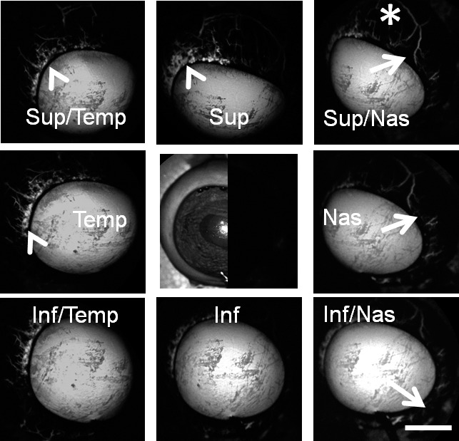
Figure 2.

Aqueous angiography with fluorescein in bovine eyes. Images from different positions were taken on a representative cow eye using fluorescein for aqueous angiography demonstrating segmental and differentially emphasized angiographic patterns. Arrowheads denoted regions of perilimbal signal, and asterisks highlighted regions of distal signal. Arrows showed areas of relatively low perilimbal signal. The central image was a composite image of cSLO infrared (left side) and preinjection background (right side) images. Sup superior; Temp, temporal; Nas, nasal; Inf, inferior. Scale bar: 1 cm.
