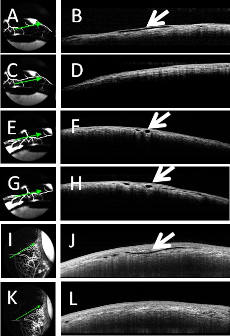
Figure 6.

Aqueous angiography and OCT in cow eyes. Aqueous angiography was conducted in cow eyes in parallel with anterior segment OCT with fluorescein (A, C, E, G) or ICG (I, K). (A, E, G, I) Angiographically-positive areas demonstrated (B, F, H, J) intrascleral lumens on OCT (arrows). However, angiographically lacking areas (C, K) were (D, L) devoid of intrascleral lumens on OCT.
