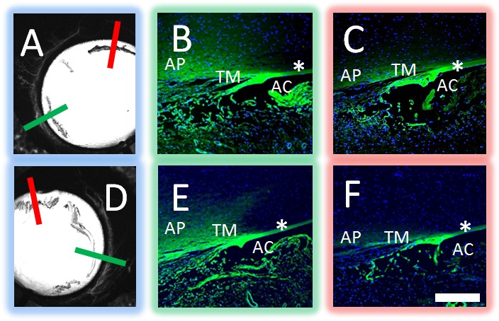
Figure 7.

Aqueous angiography localized to AHO pathways in cow eyes. Aqueous angiography was performed with 3 kD fixable fluorescent dextrans in cow eyes. Two representative eyes (A–C and D–F) are shown here. Angiographically positive (A, D; green lines) or diminished (A, D; red lines) regions were identified with aqueous angiography, marked, and prepared for paraffin sectioning. In the first eye (A–C), angiographically positive (green line in [A] corresponds to [B]) but not angiographically diminished (red line in [A] corresponds to [C]) regions showed trapping of dextrans within outflow pathways. In the second eye (D–F), angiographically positive (green line in [D] corresponds to [E]) but not angiographically lacking (red line in [D] corresponds to [F]) regions also showed trapping of dextrans within outflow pathways. Note similar degree of nonspecific fluorescence seen in strips of Descemet's membrane and iris edge in all cases (asterisks). As the fixable fluorescent dextrans were attached to nearby surfaces with fixation, nonspecific positive fluorescence lining the AC was expected after introduction of perfusion fixation. Scale bar: 100 μm.
