
Figure 8.
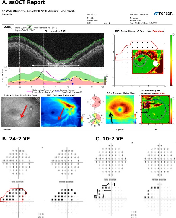
(A) Wide-field SS-OCT report for the right eye of the same patient as in Figure 7. The red arrows indicate a clear arcuate defect on the RNFL enface and RNFL thickness maps. (B, C) The 24-2 and 10-2 VFs for this eye. The contours indicate a region with abnormal points on the 24-2 (red) or 10-2 (black) VFs.