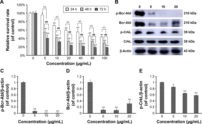
Figure 1.
Realgar NPs induced cell death and fusion protein degradation.
Notes: (A) K562 cells were seeded in 96-well plates and treated with specific concentrations of realgar NPs (0, 5, 10, 20, 40, 80, and 100 μg/mL) for 24, 48, or 72 h. The survival rate was analyzed by using CCK 8 assay at 450 nm. n=3; *P<0.05, **P<0.01, realgar NP treatment groups versus 0 μg/mL group; ##P<0.01, 48 h group versus 72 h group; ∆∆P<0.01, 48 and 72 h groups versus 24 h group. (B) K562 cells were treated with specific concentrations of realgar NPs (0, 5, 10, and 20 μg/mL) for 24 h. Bcr-Abl, p-Bcr-Abl fusion protein, CrkL, and p-CrkL protein levels were estimated by using Western blot analysis. β-Actin was used as loading control. (C) Quantification of p-Bcr-Abl/β-actin shown in (B). (D) Quantification of Bcr-Abl/β-actin shown in (B). (E) Quantification of p-CrkL/β-actin shown in (B). n=3, *P<0.05, **P<0.01, realgar NP treatment groups versus 0 μg/mL group.
Abbreviations: Abl, Abelson murine leukemia; Bcr, breakpoint cluster region; CCK-8, cell counting kit-8; NP, nanoparticle.

