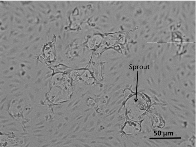
Fig. 4.
Dermal ECs in a well after stimulation with 25 ng/mL VEGF and 2 ng/mL TNF-. The circular structures form the boundaries of newly formed sprouts. This figure represents a magnification of ten times with respect to Fig. 3. One of the sprouts has been indicated by an arrow

