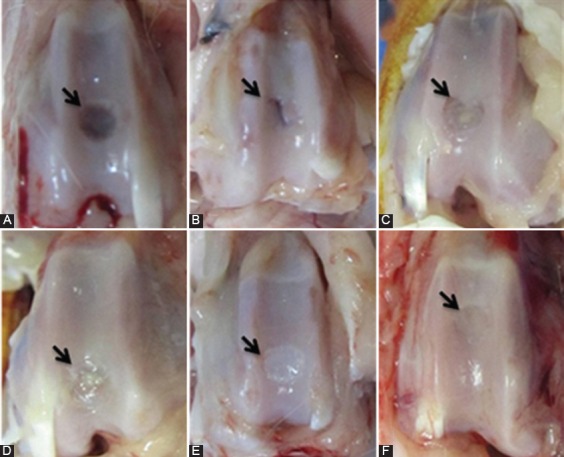
Figure 1.

Gross appearance of defects in the trochlear groove at four weeks after surgery, respectively. A) Control group: Transplantation was not performed. The defect is partially vacant and clearly noticeable from the surrounding cartilage. B) PRP group: Healing tissue is thinner than normal surrounding cartilage and shows a large fissure. C) PRF group: Repaired tissue covers defect with a minor concavity. D) Gel+SDF1 group: Repaired tissue covers the defect almost completely, but is thin and distinguishable from the surrounding cartilage. E) PRP+SDF1 group: Healing tissue covers defect with smooth white tissue and distinguishable from the surrounding cartilage. F) PRF+SDF1 group: Repaired tissue covers the defect completely and no obvious margin was notable.
