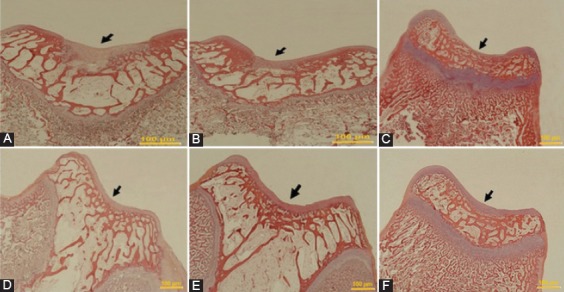
Figure 3.

The microscopic appearance of H&E staining of defects in the trochlear groove at four weeks after surgery, respectively (Original magnification=200×). A) Control group: The defect is concave and a noticeable thin layer of non-cartilaginous tissue. B) PRP group: The defect is filled by fibrous tissue with a crack. C) PRF group: The defect is covered by fibrous tissue. D) Gel+SDF1 group and (E) PRP+SDF1 group: The defects in both groups are filled with a repaired tissue, which are thinner than the normal surrounding cartilage. F) PRF+SDF1 group: The defect is entirely filled by the repaired tissue, resembling the healthy cartilage surrounding the tissue.
