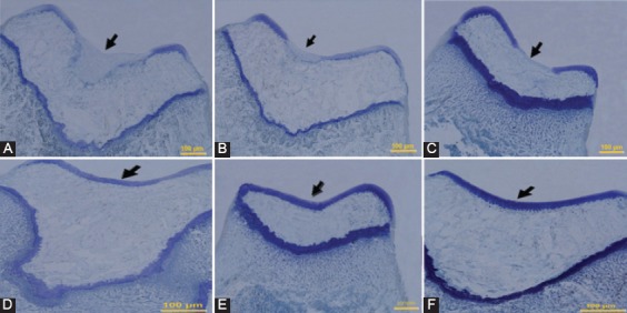
Figure 4.

Histological appearance of Toluidine blue staining of defects in the trochlear groove at four weeks after surgery, respectively (Original magnification=200×). A) Control group: The defect is filled by no cartilage matrix. B) PRP group, and C) PRF group: The defects in both groups have poor staining fibrocartilagous tissues and poorly organized ECM. D) Gel+SDF1 group, and E) PRP+SDF1 group: The repaired tissues show a good staining in both groups. F) PRF+SDF1 group: The defect has an intense staining.
