
Fig. 5.
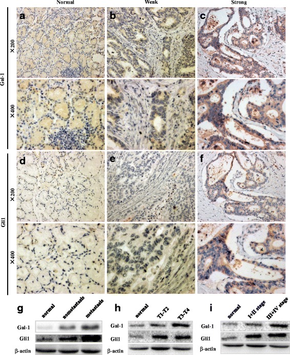
Immunohistochemical staining of Gal-1 and Gli1 in GC sections. Expression of Gal-1 is low in normal tissues (a), while staining ranges from weak (b) to strong (c) in GC tissues. Likewise, Gli1 shows low expression in normal tissues (d), while expression ranges from low to high in GC tissues. Western blot analysis of Gal-1 and Gli1 expression in GC tissues showing that expression is significantly correlated with lymph node metastasis (g), the depth of tumor invasion (h), and TNM stage (i)