Fig. 2.

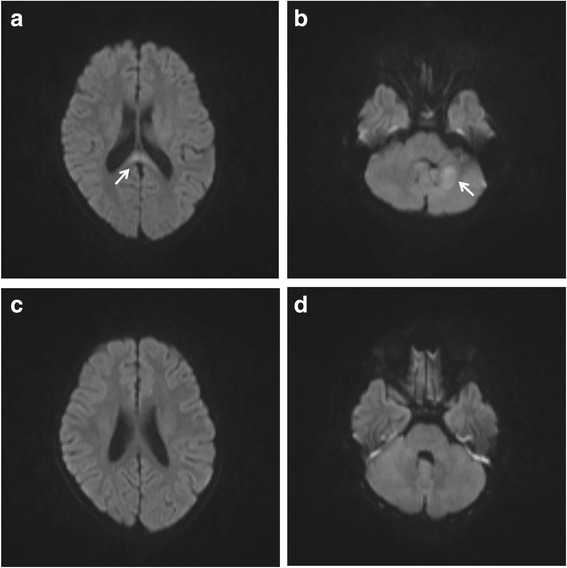
The brain MRI in case 2. The brain MRI on day 5 revealed high intensity lesions (arrows) in the SCC (a) and the left cerebellum (b) on diffusion-weighted images, which disappeared on day 12 (c and d)

The brain MRI in case 2. The brain MRI on day 5 revealed high intensity lesions (arrows) in the SCC (a) and the left cerebellum (b) on diffusion-weighted images, which disappeared on day 12 (c and d)