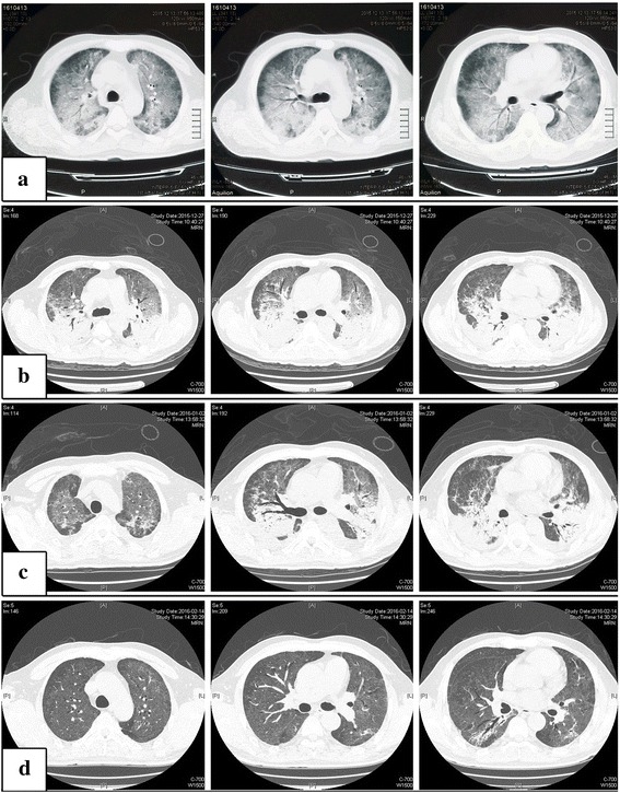
Fig. 2.

High resolution CT scans of the chest at the levels of aortic arch, root of ascending aorta and pulmonary arteries from left to right, performed from top to down on days 1 (a: December 2015), 14 (b: December 2015), 20 (c: January 2016) and 90 (d: February 2016). Bilateral lung infiltrates with ground-glass attenuation (a). Bilateral infiltrates and dense consolidations aggravated (b). Minimal absorption compared to day 14 (c). Dense consolidations were significantly absorbed (d)
