Figure 3.
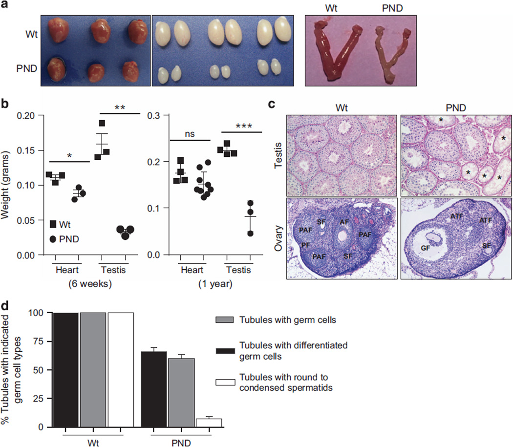
Morphological differences in mouse tissues. (a) Representative images showing comparative size of heart, testis and uterine horns in wild-type and Mdm2PND/PND mice. (b) Comparative analysis of heart and testis weights from 6 weeks (left) and 1-year-old (right) wild-type and Mdm2PND/PND mice.±s.e.m., *P < 0.05, ** P < 0.01, ***P < 0.001. ns, non significant. (c) Representative periodic acid–Schiff (PAS)-hematoxylin-stained cross-sections of testis (20×) and hematoxylin and eosin (H&E)-stained sections of ovary (5×) from wild-type and Mdm2PND/PND mice. (d) Graphical representation of percentage of seminiferous tubule sections containing germ cells of different types in wild-type and Mdm2PND/PND mice testes. *Seminiferous tubules without germ cells; AF, antral follicle; ATF, atreal follicle; GF, Graffian follicle; PAF, preantral follicle; PF, primary follicle; SF, secondary follicle.

