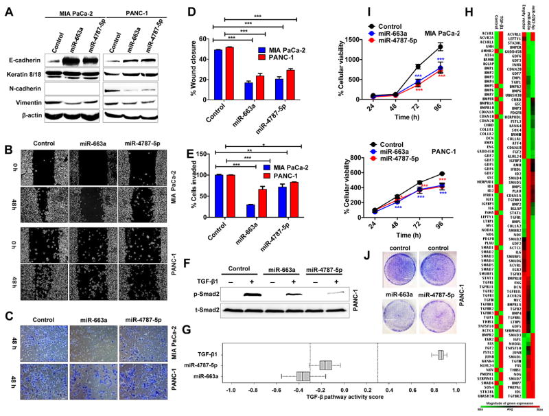
Figure 6. MiR-663a and miR-4787-5p resists EMT features and slows cellular growth in pancreatic cancer cells.
A. MiR-663a and miR-4787-5p increased epithelial markers and decreased mesenchymal markers. Whole cell lysates (50 μg) of cells overexpressing miRNAs were subjected to Western blotting for EMT markers with β-actin as a loading control. B & D. MiR-663a and miR-4787-5p decreased cell migration. Representative images of cells subjected to a scratch wound assay (B) and quantification of wound closure measurements (D). Original magnification X4. C & E. MiR-663a and miR-4787-5p decreased cell invasion. F. MiR-663a and miR-4787-5p reduced p-Smad2 levels. Whole cells lysates of TGF-β1 (10 ng/mL; 60 minutes) treated cells subjected to Western blotting to detect total and p-Smad2. G & H. MiRNAs decreased expression of TGF-β1-responsive genes and attenuated TGF-β1 signaling pathway. Total RNA from cells subjected to a RT2-PCR profiler array. Heat maps showing transcript level changes of TGF-β-responsive genes (H) and overall TGF-β pathway activity scores depicted (G). I & J. MiR-663a and miR-4787-5p reduced cellular proliferation as determined by MTT (I) and colony formation (J) assays. Points, mean of triplicate; bars, SD. n=3

