Figure 1.

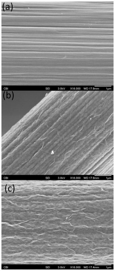
Scanning electron microscopy images of a bare CFE (a), a 25 s PEDOT/GO coated CFE (b) and a 100 s PEDOT/GO coated CFE (c). All three images display an 18,000X magnification of the electrode surfaces. The scale bars represent 1 μm.

Scanning electron microscopy images of a bare CFE (a), a 25 s PEDOT/GO coated CFE (b) and a 100 s PEDOT/GO coated CFE (c). All three images display an 18,000X magnification of the electrode surfaces. The scale bars represent 1 μm.