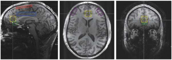
Figure 2. Proposed Regions of Interest to Identify Alterations in Glu/GABA and Abnormalities in Functional Connectivity.
Sagittal, axial, and coronal viewpoints showing the medial prefrontal cortex (mPFC) in yellow. Sagittal viewpoint showing the fronto-parietal cortical region in red and the dorsal anterior cingulate cortex (dACC) in blue. Sagittal and coronal viewpoints showing the subgenual anterior cingulate cortex (sgACC) in green. Axial viewpoint showing the dorsolateral prefrontal cortex (DLPFC) in purple. Hippocampus and amygdala not shown in figure.
*Modified with permission: http://onlinelibrary.wiley.com/doi/10.1002/jmri.24970/full#jmri24970-fig-0001

