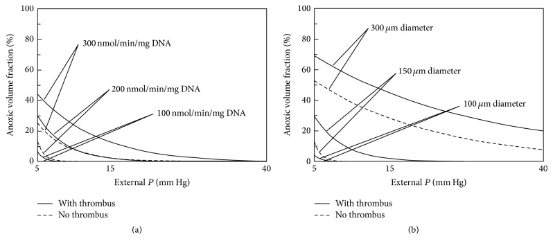
Figure 5.
Graphs depicting a summary of model results for calculation of anoxic volume fraction [AVF (%)] with respect to external blood P (P ext), islet fractional viability (OCR/DNA), and diameter (2 · R) and with or without thrombus formation (δ = 100 μm). The graph on the left (a) illustrates the change in AVF for an islet of average diameter (150 μm) for the 3 OCR/DNA values. The graph on the right (b) illustrates the change in AVF in an islet with average OCR/DNA (200 nmol/min/mg DNA) for 3 islet diameter values. AVF is defined as the region of the islet that is anoxic, occurring below a critical P (P C) of 0.1 mm Hg. AVF, anoxic volume fraction; δ, thickness of the thrombus; OCR, oxygen consumption rate; P, oxygen partial pressure; P C, critical P for viability; P ext, external blood P; R, islet radius.

