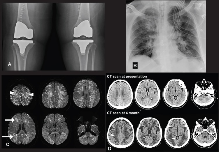
Figure 1.

Radiological findings A) Postoperative knee radiograph is demonstrating metallic prosthesis in both knees, B) An immediate postoperative chest radiograph is showing acute interval development of bilateral infiltrated most pronounced in the respective peri-hilar regions and right upper lobe, C) Selected axial diffusion-weighted MRI images are demonstrating multiple small foci of diffusion restriction (confirmed by apparent diffusion coefficient maps) with a near symmetrical distribution predominantly regarding to the watershed territories (arrows). Some of the high bilateral parietal and frontal lesions were cortical based (arrow heads), D) Selected axial non-contrast enhanced CT scans of the brain at presentation and 4-month follow-up. The follow-up images (below) show extensive low attenuation areas in a watershed territory associated with accelerated central brain atrophy (arrow heads).
